EEG (Electroencephalography)
EEG is a non-invasive technique for measuring the electrical activity of the brain. It is measured as a time-varying voltage difference between electrodes placed on a person’s scalp. EEG generally monitors the activity of the neurons in the cerebral cortex as it is closest to the electrodes. The electric potential generated by an individual neuron is far too small to be picked up by EEG and hence it measures the synchronous activity of thousands or millions of neurons. The measured voltage difference between the electrodes is only in the range of microvolts and needs amplification for the recordings to be made. The contribution from the neurons further away from the scalp is even weaker and hence EEG can not measure deeper brain structures. Nonetheless, the cerebral cortex is probably the most interesting part of the brain, which makes us human, allowing us to think, feel, and experience the world around us.
Cortex topography
The cortex can be divided into different regions, called lobes, each responsible for various brain functions. Knowing cortex topography is important when choosing the placement of electrodes for EEG measurements when performing experiments on developing EEG-based systems.
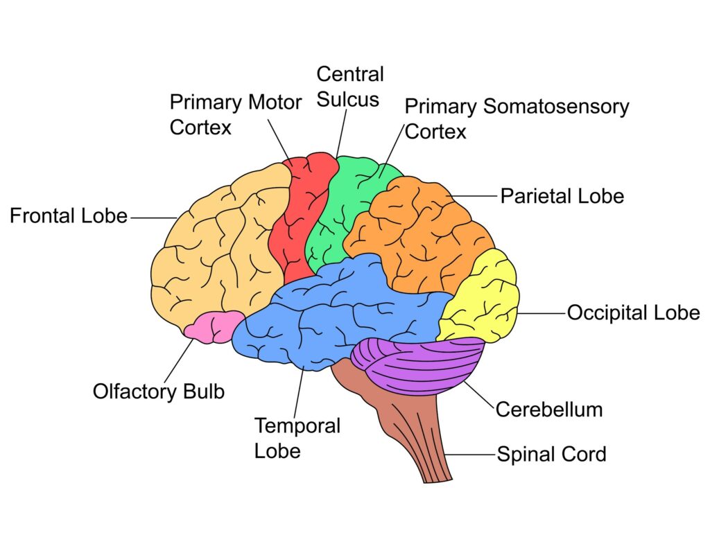
For example, the motor cortex is responsible for motor commands. Changes in activity can be observed there when a person intends and/or makes a movement. Likewise, sensorimotor cortex activity varies when a person’s sensory nerves are activated by the external environment. The occipital cortex region is responsible for processing visual information and so on.
Brainwaves
Brainwaves are electrical oscillations or rhythms observed in EEG recordings. Various oscillations can reflect the state of mind of the person such as tiredness, relaxation, concentration, etc. There are associated oscillations with different sleep stages so it can be used to study and determine these. Brainwaves are categorized into different frequency bands, each associated with specific mental states, activities, or cognitive functions.
Delta Waves (0.5 – 4 Hz). Associated with deep sleep, unconsciousness, and certain abnormal brain states.
Theta Waves (4 – 8 Hz). Associated with dreaming, creativity, meditation, and deep relaxation.
Alpha Waves (8 – 13 Hz). Associated with wakeful relaxation, daydreaming, and a calm mental state.
Beta Waves (13 – 30 Hz). Associated with normal waking consciousness, concentration, and cognitive tasks.
Gamma Waves (30 – 100 Hz). Associated with higher cognitive functions, perception, and consciousness.
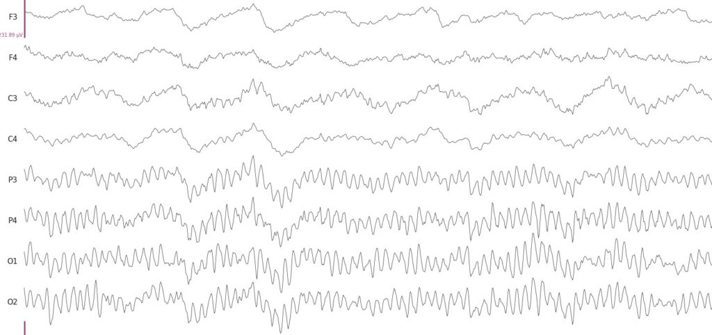
An example of the so-called alpha waves is shown in the figure below. The alpha rhythm can be observed over the occipital cortex region when a person closes their eyes and/or their visual activity is at rest.
Event Related Potentials (ERP)
EEG is used extensively to measure Event-Related Potentials (ERP). Event-Related Potential is neural activity that is time-locked to specific sensory, motor, or cognitive events. In essence, it is the brain’s response to certain stimuli or events.
ERPs are composed of several different components, each of which is associated with a specific cognitive process. Some of the most common ERP components include:
P300: The P300 is a positive ERP component that is typically elicited by the presentation of target stimuli. It is thought to reflect the brain’s response to novelty, surprise, or expectation.
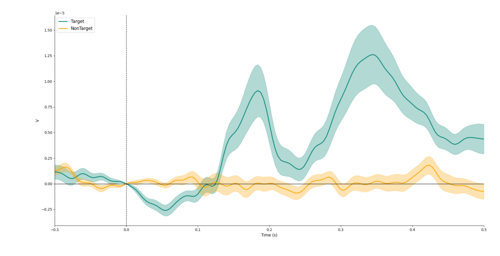
N170: The N170 is a negative ERP component that is typically elicited by the presentation of faces. It is thought to reflect the brain’s early processing of facial features. This can be seen as a negative deflection in the figure above.
LPP: The late positive potential (LPP) is a positive ERP component that is typically elicited by auditory stimuli. It is thought to reflect the brain’s sustained attention to sounds.
ERPs are a powerful tool for studying the brain’s electrical activity in response to specific events. They provide valuable insights into the temporal dynamics of cognitive processes and have widespread applications in both basic and applied neuroscience research.
Artifacts
EEG recordings capture not only the neural activity of the brain but also other electrical signals that come from more powerful sources. These are called artifacts and may hinder visual interpretation of the signals or can even be mistaken for actual EEG activity. Typically, it is sought to either minimize the artifacts or remove them altogether from EEG recordings with various processing techniques to have a clean EEG signal. The main sources of these artifacts are:
Eye blinks and movements. These are high-amplitude signals observed in EEG recordings, particularly over the forehead and frontal lobes. They can be easily detected and there are now many algorithms that try to address their automated removal from EEG signals such as ASR (link).
Muscle activity. Movement of the neck, jaw, and tongue generates electrical signals due to muscle contractions that are relatively large and hence are picked up by EEG electrodes that are in a nearby location. Electrical signals associated with muscle activity are wideband and fall in the range of Beta and Gamma waves making their removal a challenging task.
Cardiac activity. The potential for cardiac activity can be also picked up in the EEG recording. This is particularly visible if an electrode is placed on some major blood vessel. Cardiac signal artifacts can be removed, for example, by simultaneously recording an electrocardiogram (ECG) and using this information for the removal of the artifacts from EEG signals.
Environmental noise sources. This is usually caused by electrical equipment powered by the main electricity grid and manifests as 50Hz or 60Hz frequency noise in EEG recordings. This can be minimized either by removing the sources from the recording environment or using active electrodes (link), better shielding of measuring equipment, better electrode contact with a scalp, etc. If the mains noise is not extremely large, it can be removed from the signals with conventional filtering techniques.
Applications
There are many applications for EEG measurements and the use cases will keep growing as the accessibility of EEG devices keeps increasing. The primary applications for EEG are still in the fields of medicine and research but it will likely grow into new emerging applications such as Neuromarketing.
Medical. EEG recordings are used to diagnose epilepsy, sleep disorders, stroke, encephalitis, and other brain disorders. It can be used in treatment as well when some parameters of EEG signals are used as feedback to a person, known as neurofeedback. It has been shown, for example, to help patients recover motor skills more efficiently after a stroke.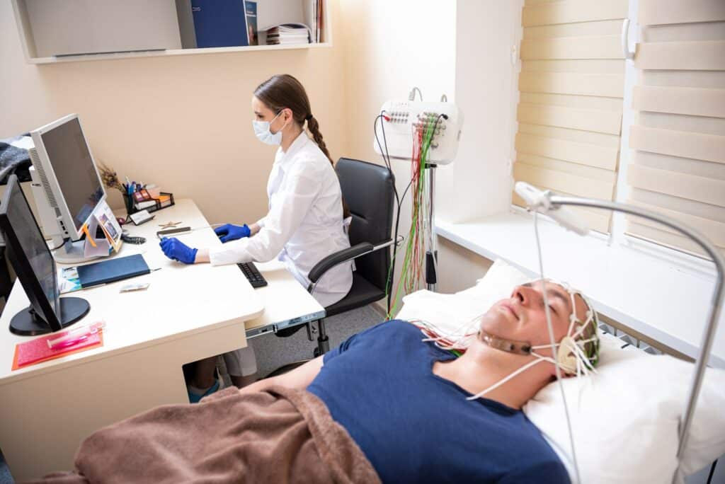
Research. EEG is extensively used as a tool in cognitive neuroscience, clinical neuroscience, psychology, neurolinguistics, and others. Researchers are using and studying, for example, the aforementioned ERPs to gain an understanding of cognitive functions, attention, and decision-making mechanisms. Various potential biomarkers are identified as potential indicators for neurological and psychiatric conditions aiding early diagnosis and treatment monitoring. EEG is also used to investigate neural mechanisms involved in memory formation consolidation and retrieval. It provides insights into neural plasticity and learning processes.
Brain-Computer Interface (BCI). As a non-invasive and accessible method, EEG is quite often used to measure brain activity in BCI systems. BCI systems aim to transfer the intention/command of a person to a computer or other device. Most popular BCI approaches are based on motor imagery, P300 Speller, SSVEP, and others. Read more on this in the BCI tutorial (link)
Neuromarketing. While traditionally gaze tracking is the major measurement technique used in neuromarketing, EEG measurements are also becoming more popular as they provide additional insights into how we respond to products, brands, advertisements, etc. EEG is used to measure the user’s attention, engagement, memory, and recall. It helps improve user experience with certain products, website and app designs, and others.
Measurement setup
Conventional EEG recording is made by placing electrodes on the scalp and applying conductive gel in between to establish electrical contact. The scalp is usually also prepared with light abrasion to reduce impedance due to dead skin cells.
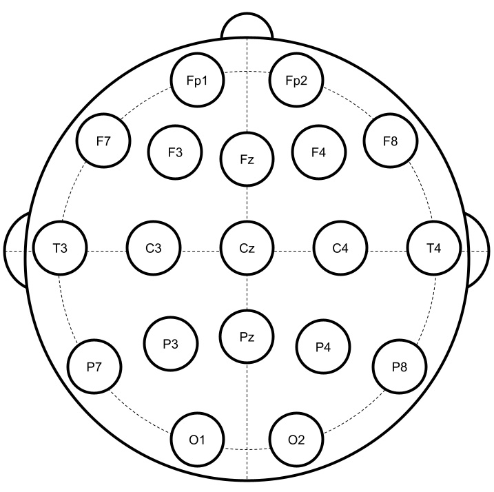
10/20 System. It is a standardized approach used for the notation of electrode placement. It ensures that electrodes with the same name will be placed at the same relative location concerning other electrodes irrespective of the head size or number of electrodes used by the measuring system. An example of 19 electrode placement and their naming is shown in the Figure below. The standard notation helps clinicians, researchers, and others to easily compare their measurements knowing exactly the topographic location over which the measurement was made.
Referential montage. As mentioned previously EEG measures the voltage difference between two electrodes. In sequential montage, the EEG measurements are made between pairs of electrodes, typically placed relatively close by. A more popular approach though, is to use a common reference electrode, where a voltage difference is measured between any electrode and this reference electrode. However, the placement of the reference electrode is not standardized and some setups place the reference electrode on the ear, some on the forehead, or some other location yielding measurements directly incomparable between different setups. Nonetheless, the signals can be re-calculated in postprocessing to use an average reference or REST (reference electrode standardization technique) standardizing these common reference EEG recordings.
Dry-contact electrodes. Whilst traditional EEG setup involves the application of conductive gel or paste to establish electrode contact, dry-contact electrodes that exclude the need for gel are becoming more popular, reducing the setup time and improving general comfort. This is particularly important for measurements outside of the laboratory in real-life environments and non-medical or research use cases. It is worth noting that BrainAccess products use innovative dry-contact EEG electrodes that conform to the shape of the head and make them more comfortable to wear than traditional dry-contact electrodes. However, dry-contact electrodes are less stable than gel-based electrodes requiring more post-processing procedures to ensure good quality recordings.
Passive and active electrodes. There are two types of electrodes in terms of signal amplification that are used in EEG recordings. Passive electrodes, where the electrodes are connected with cables to the measuring device where the signal amplification and digitization are performed. Active electrodes have preamplifiers built into the electrode that amplify the EEG signal before sending it to the measuring device. This helps reduce the environmental electrical noise picked up by the cables which can lower the quality of the measured EEG recordings. However, active electrodes add more complexity and are more costly when compared to passive electrodes.
